GO TO INDEX
CHAPTER 2: HOMEOSTASIS
Questions And Answers
Q.1: What is an internal environment of an organism?Ans: Internal Environment of an Organism:
The internal conditions of an organism are referred as its internal environment; it includes H2O quantity, different solutes, temperature, etc. For proper metabolic functions, the body requires all these conditions at a particular level.
Q.2: Why should the internal environment of living organisms remain constant? Give an example?
Ans: The internal conditions of an organism are referred as its internal environment; it includes H2O quantity, different solutes, temperature, etc. The internal environment of organisms remain constant for proper metabolic functions; the body requires all these conditions at a particular level.
Lets take the example of temperature, the temperature of external environment is continuously change during day but the enzymes work within a certain range of temperature therefore, the living organisms maintain internal temperature within this range. Organisms maintain internal condition by feedback mechanism.
Q.3: Define homeostasis? What are the important aspects of homeostasis OR Briefly describe the adaptation of plants in different conditions?
Ans: Homeostasis:
Homeo means same and stasis means state or condition.
Homeostasis is set of metabolism which maintain internal environment of an organism within suitable limits.
Important Aspects of Homeostasis OR The Adaptation Of Plants In Different Conditions:
There are three important aspects of homeostasis.
- a. Osmoregulation:
It is the maintenance of internal water and salt conditions by osmosis. - b. Thermoregulation:
It is the maintenance of temperature within suitable limits where enzymes can work properly (optimally). - c. Excretion:
It is the process where metabolic toxic waste or excess metabolic substances (i.e NH3, urea or uric acid, gums, latex etc.) from body are removed.
Q.4: Define feedback system? Also draw a block diagram to show that how body maintains homeostasis by the feedback mechanism?
Ans: FEEDBACK SYSTEM:
The check and balance system in a body is called feedback system. In a feedback system three organs are involved.
- Receptor:
The organ which receive any change in the internal environment of the body. The receptors transfer messages to CNS. e.g. 5 sense organs. - Central Nervous System:
It is the major command and control center to which stimuli are reported by receptors and decisions are made and conveyed to effectors. Such as brain. - Effector:
The central nervous system sends the message to a particular organ. Response is exhibited by these organs like muscles, glands etc., are termed as effectors.

Q.5: Describe excretion or storage of CO2 in plants?
Ans: Excretion or Storage of CO2 In Plants:
At daytime plant perform photosynthesis in green cells and respiration in all living cells. The CO2 produced in respiration utilized in photosynthesis. When rate of photosynthesis will be higher than respiration the plant gets extra CO2 from air and release extra O2 in air through stomata.
At night plant only perform respiration only CO2 is produced which is removed by the process of diffusion through body surface. The green parts perform these gases exchange through stomata while non green parts perform this gaseous exchange through body surface.
Q.6: Define Transpiration and Guttation OR Describe how extra water is removed from plants?
Ans: Removal Of Extra Water In Plants:
The plant store large amount of water this water can be removed from plant in two ways i.e.
(a) Transpiration
(b) Guttation
Transpiration:
It is the removal of water in the form of vapours from aerial part of plant. It occurs only at day time.
Plants modify their leaves size, structure and structure of stomata to control the rate of transpiration.
Guttation:
It is the removal of water in the form of liquid from the margin of leaves through special pores, hydathodes. It only occurs at night when water pressure is high in leaves and low temperature environment is present.

Q.7: Differentiate between transpiration and guttation.
Ans: Difference Between Transpiration And Guttation
| S.NO. | Transpiration | Guttation |
|---|---|---|
| 1. | It usually occurs during the day. | It usually occurs at night or early in the morning. |
| 2. | Water is given out in the form of water vapours. | Water is given out in the form of a liquid. |
| 3. | Several factors are responsible for this process. | This primarily occurs due to higher root pressure. |
| 4. | Water comes out in the pure form. | Water droplets consist of various organic and inorganic salts dissolved in them. So it is not pure. |
| 5. | It occurs through stomata, lenticels or cuticles. | It occurs through hydathodes only. |
| 6. | It is a controlled phenomenon. | It is an uncontrolled phenomenon. |
Q.8: How metabolic toxic waste or excess metabolic substances are removed from plant body?
Ans: Removal Of metabolic toxic waste:
Plants produce some secondary products like latex, resin and gum. These secondary products are insoluble, harmless compounds. Some plants produce special types of gums for example Neem or keeker etc. The extra amounts of these are removed from special pores called lenticels. The coniferous plants produce resins like material while the rubber plants produce latex which removes from scare like openings. Some of these carnivorous plants and okra produce mucilaginous material to capture insects.
Q.9: Describe types of plants on the basis of salt and water quantity in different conditions?
Ans: Types Of Plants:
The plants grow in different conditions of water and salts, on the basis of water and salt quantity there are four type of plants
- Hydrophyte
- Halophyte
- Mesophyte
- Xerophyte
Hydrophytes (hydro = water; phyta = plants):
The group of plants which grows in fresh water are called hydrophyte. They live completely or partially in fresh water so called totally or partially submerged plants. They adapt themselves for removal of excess water which can enter in this condition.
Characteristic of Hydrophyte
- The plant do not have layer of cuticle.
- These plants do not contain roots or poorly developed roots.
- They have broad leaves if partially submerged and have stomata in the upper surface epidermis (to take part in transpiration) e.g. water lily.
- They may have thin and spongy tissues in leaves and stem in totally submerged plant e.g. Hydrilla.
- Examples are Hydrilla, Lotus, Lily plant
Halophytes (Halos= salt):
The group of plants which grows in sea marshes or salty water are called halophyte. In salty condition water moves outside the cell which is not suitable for plants. To move water from outside to inside the plant develop following characters.
Characteristic of Halophyte
- They absorb water from the soil, which has higher salt concentration and low water potential.
- They actively absorb salt into their roots.
- Plants develop salt glands where plant store salts by taking it through active transport.
- Plants oppose salt to move outside from vacuole.
- Some salt accumulated at surface of leaf which attracts water from air.
- Some halophytes absorb humidity by leave.
- Examples are Glasswort, Cord grass
Mesophyte:
The group of plants which grows in well watered soil (or moderate water containing soil) are called Mesophyte.
Characteristics of Mesophyte:
- They have developed root system which does not grow very deep.
- Their body is covered by a layer called cuticle.
- They contain stomata for evaporation of extra water.
- They have moderate sized leaves.
- Some Mesophyte excrete out water in the form of drops ( guttation)
Xerophytes:
The group of plants which grows in soil of low water quantity or in dry places are called Xerophyte. They grow in desert or steep slopes or at high altitude. To conserve water and absorb proper amount of H2O they develop following characters.
Characteristic of Xerophyte:
- They have vertically growing deep root system to absorb proper amount of water.
- Some plant have horizontal root on the surface to absorb rain water rapidly.
- They possess thick waxy cuticles over epidermis to conserve water.
- They have short sized leaves or some plant leaves are modified into spine to prevent transpiration by reducing the number of stomata.
- Some xerophytes has special parenchyma cells in stem, where they store water, this makes the stem soft, wet and juicy called succulent organs .g. cacti.
- Examples are Cactus, Euphorbia.

Q.10: Discuss Osmoregulation in animals in detail.
Ans: Osmoregulation In Animals:
Animals live in aquatic and terrestrial habitat. According to their environment their cells require critical balance of water and solutes. Water continuously leaves and enters the cells with solutes to keep the water and solute in constant quantity to ensure smooth metabolic functions.
Osmoregulation in Aquatic Environment:
Concentration of salt in water defines the aquatic conditions. The water which contains very low amount of salt is called fresh water and the water which contains high salt is called marine water. Animals osmoregulate differently in both waters.
i. Osmoregulation in fresh water:
Fresh water animals have hypertonic conditions inside their body or cells so they are always facing the problem of flooding of H20 and loss of salts. These animals are further classified in two groups.
-
Unicellular:
* They pump out excess water by contractile vacuole.
* E.g. Amoeba, Paramecium etc. - Multicellular:
* They pump out excess water by producing dilute urine.
* Loss of salt is compensated by active uptake of salt by gills and skin as well as use of salt containing food.
ii. Osmoregulation in marine animals
Marine animals have mostly hypotonic conditions (low salt) inside the body but some of them develop hypertonic (high salt) or isotonic (same salt condition) by metabolism. These animals are further classified as:
-
Bony Fish:
* Have low salt inside the body.
* Actively get sea water and have salt glands to increase the salt and desalination.
* Produce concentrated urine. - Cartilaginous Fish:
* Have high salt by storing urea inside.
* Eat food which contain nitrogenous compound i.e. meat. - Osmoconformer:
* Have equal amount of salt.
* These animals do not require any activity to adjust their internal osmotic condition. i.e. unicellular.
iii. Osmoregulation in terrestrial condition:
In land animals excretion of water take place through body surface which leads to dehydration so they have developed number of strategies to maintain Osmoregulation.
Only arthropods, some molluscs, reptiles, birds and mammals can survive in this habitat because:
- Their bodies are covered by exoskeleton or thick skin, which prevent loss of water.
- They conserve water by reabsorption in kidneys and rectum.
- Some of them can produce water from fats catabolism with the help of peroxisomes i.e. camel, kangaroos.
- Continuously drinking of water or using liquid food.
Q.11: Define and explain Excretion? Also describe excretion in plants and animals?
Ans: Definition Of Excretion:
The removal of harmful substances produced in the metabolic process (nitrogenous metabolic waste) from the body is called Excretion.
Explanation:
During metabolism living organisms catabolize protein and other nitrogen containing compounds which produce some toxic nitrogenous compound. These toxic compounds are mainly NH3 or urea or uric acid generally called nitrogenous waste. If these compound retain in the body and accumulate, they can damage the cells or organs therefore they must be removed from the body. The removal of these nitrogenous metabolic wastes is called excretion.
Excretion in Plant:
In plant rate of catabolic process is very slow and waste products are produced in less amount. They are used again in their anabolic process.
Plants are autotrophs, initially they produce carbohydrates as primary products. Carbohydrate is catabolized to produce CO2 and H2O. The CO2 reutilized in photosynthesis and H2O is not a toxic compound. As autotrophs they synthesize variety of compounds, so the waste products of one reaction are utilized in other metabolic reactions as reactant and consumed. Excess amount of waste materials is remove as:
- Excess water is removed by transpiration and guttation.
- Excess oxygen and carbon dioxide are removed through photosynthesis and respiration.
- Excess amount of ion are deposit into dead cell of plant body such as bark.
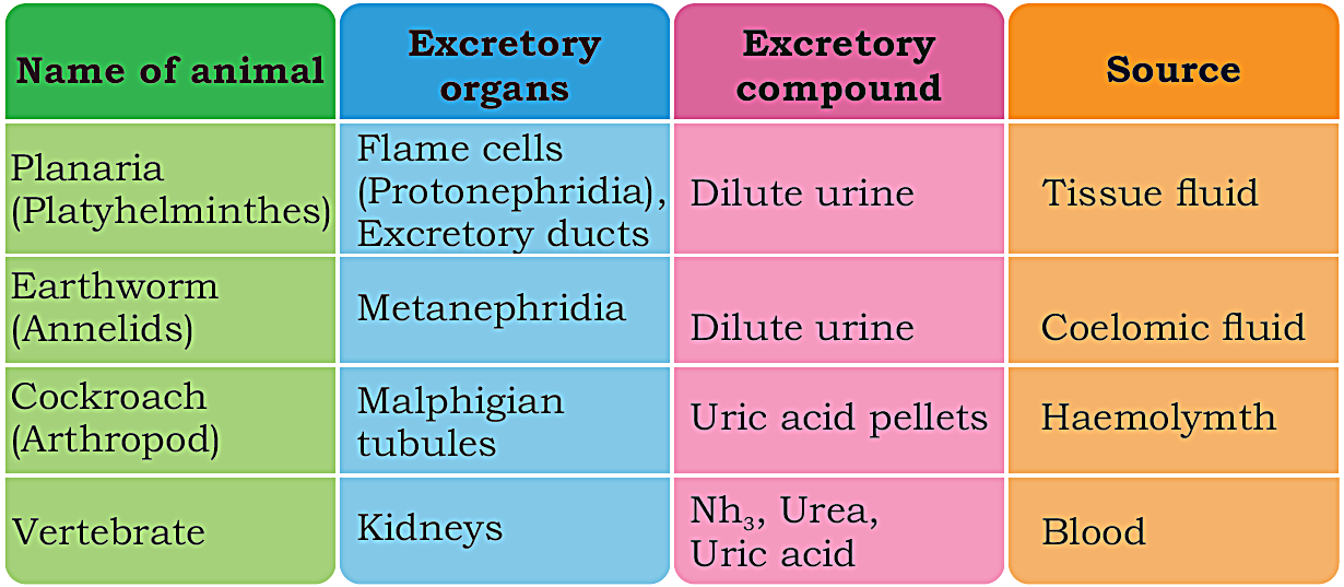
Excretion in Animal:
In animal removal of nitrogenous waste from the body is very essential. Animal have particular organ to excrete out nitrogenous waste. These organs are called excretory organs. There are different types of wastes in animals for which different excretory organs are used. The details are as follows:
Q.12: What are the main organs of human homeostasis system? OR Explain Homeostasis in man.
Ans: HOMEOSTASIS IN MAN
Humans has well developed homeostasis system. The main organs which involved in homeostasis are:
- Skin
- Lungs
- Kidneys
(i) Skin:
The Skin is considered as the largest organ of the body, basically functions as a protective organ as the first line of defence but it also works efficiently as a homeostatic organ by maintaining temperature, water and salt. It excretes water, salt, and urea from the body through sweat.
(ii) Lungs:
Lungs are responsible for maintaining levels of O2 and CO2 in the blood, body fluid and cells in order to maintain the rate of respiration and continuous flow of energy.
(iii) Kidneys:
Kidneys are called filters of the body fluids, they maintain internal water by removing excessive water, also maintain urea, uric acids, creatinine and other waste by excreting them through urine.
Q.13: Explain the structure of Human skin in detail with labelled diagram?
Ans: STRUCTURE OF SKIN:
Human skin is consists of three layers ,namely:
- Epidermis,
- Dermis and
- Hypodermis.
- Epidermis is the outer most layer which is made up of flat, dead cells containing keratin protein.
- This layer does not contain blood vessels.
- It is an impermeable to water and thus prevents water loss from the body.
- It also works as protective layer by preventing the entry of micro organisms.
- It also has sweat pores.
- Hairs are also emerge (come out) from epidermis.
Dermis:
Dermis is the layer present between epidermis and hypodermis, it contains many different structures i.e.:
- Dermis has nerves ending receptors to detect temperature change, pain, pressure etc.
- It also contains sweat glands which secrete sweat on the surface to maintain temperature and also secrete urea, water and salt.
- A network of arterioles is also present in the form of network, which are involved in temperature regulation.
- The dermis also contains hair follicle and sebaceous glands which secrete oily sebum.
Hypodermis:
- It is the inner most layer of skin.
- It contains fats which act as insulation against loss of heat.
- It also stores energy.

Q.14: Discuss the role of skin in regulating body temperature Or How skin works as thermoregulatory organ?
Role of Skin in Regulating Body Temperature OR Skin Works As Thermoregulatory Organ:
The skin is the organ which help in regulating body temperature .when the receptors in skin detects change in body temperature from set point (set point of human is 37 ℃) i.e. increase or decrease, and then Receptor send nerve impulse to brain. It occurs by feedback mechanism to correct the temperature.
In Hot condition If Body Temperature Start Rise: (Summer)
- Production of sweat:
The sweat gland starts to produce and secrete sweat. The sweat accumulates at the surface of skin and evaporates with heat energy so the body feeling cool. - Laying down of hairs:
In hot condition, muscles which are attached with hair relax. It allows the hair to lie flat against surface of the skin. - Vasodilation:
Arterioles found in the form of network in dermis, dilate (become wide) which increasing the flow of blood as well as it brings the blood vessels near the surface of skin which allows more heat loss. This process of vessel dilation is called vasodilation.
In Cold Condition When Body Temperature Starts Decreasing: (Winter)
In cold condition when the body temperature starts decreasing the skin maintains the temperature by following actions:
- Erection of hairs:
The muscles contract pulling the hairs upright and trapping a layer of insulating air next to skin. Now it is not very much effective in human. - Vasocontraction:
Narrowing of blood arterioles of dermis occurs which reduces the blood flow in capillaries of skin so less heat is lost. - Decrease in sweat production:
The sweat gland stops to produce and secrete sweat, so it prevent from energy loss. - Increase in metabolic rate:
In cold conditions the rate of metabolism in the organs increases generating more heat which is distributed around the body in the blood stream. It prevents loss through the adipose tissue in hypodermic which work as an insulation layer.

Q.15: Explain and discuss the role of lungs to keep the CO2 concentration low to certain level.
Ans: Role Of Lungs To Keep The CO2 Concentration Low To Certain Level:
Lungs play a vital role in keeping the CO2 concentration low to a certain level through the following steps:
- Tissues / cells produce a large amount of CO2 produced during the aerobic respiration.
- As blood passes through tissues via blood capillaries, this CO2 diffused into the blood, where it reacts with water forms carbonic acid. This reaction takes place by an enzyme called carbonic anhydrase present in R.B.C.
- The carbonic acid dissociate into H+ and bicarbonate ions. HCO3-1 ions.
- The level of H+ in blood is continuously monitored by special detectors (receptor) carotid bodies and aortic bodies.
- Most of the bicarbonate ions diffuse out from R.B.C to blood plasma.
- A small amount of CO2 is also carried and dissolved in R.B.C when the blood reaches lungs these bicarbonate ions diffuse back into RBC where again converted into carbonic acid then into CO2.
- The CO2 diffuses out of the blood capillaries and into alveoli, from where it is expelled out when breathing out.
- If the CO2 level increases in blood, pH of blood start increasing so that the receptor send a message to the control centre which ultimately increase the breathing rate to expel out the CO 2 efficiently.

Q.16: What is the role of the kidney in controlling blood composition?
Ans: Role Of The Kidney In Controlling Blood Composition:
Blood is the fluid having cells. In plasma, it contains a high amount of H2O and some solutes like Na+, Cl-, Ca++, K+ etc. with nitrogenous waste. Liver continuously produces urea and NH3 by breaking amino acid, we continuously take different solute ions in our food like Na+, Ca++, K+ etc. the concentration of H2O, solute and nitrogenous waste are maintained by kidney through process of filtration and reabsorption.
Q.17: What do you know about urinary system in man? OR Describe the urinary system of man with the help of diagram?
Ans: Urinary System In Man
Urinary system of man consist of:
- A pair of kidney
- A pair of ureters
- A urinary bladder
- A urethra
Kidneys:
- Kidneys are reddish-brown bean shaped organs, situated at the dorsal side of the abdominal cavity on either side of the vertebral column.
- The kidneys lie above the waistline.
- Each kidney has an area in the center of concave surface which faces the vertebral column; this area is called hillus.
- The renal artery, renal vein, nerve and ureter are connected to each kidney at the hillus.
- They are covered by a membrane are called peritoneum.
Ureter:
- The ureter is a narrow tube which connects the kidney to the urinary bladder.
- Urine passes through ureter to the urinary bladder.
Urinary Bladder:
- The urinary bladder is a thin walled muscular pear shaped bag situated towards the bottom of abdominal cavity in front of the rectum which stores urine.
Urethra:
- The urethra is a tube which comes out from the urinary bladder, runs down and opens outside the body through urinary opening (Anus).
- It passes urine from bladder to outside the body.

OR

Q.18: Explain the external an internal structure and function of kidneys in detail with labelled diagram.
Ans: External Structure Of Kidney:
- Kidneys are reddish-brown bean shaped organs, situated at the dorsal side of the abdominal cavity on either side of the vertebral column.
- The kidneys lie above the waistline.
- Each kidney has an area in the center of concave surface which faces the vertebral column; this area is called hillus.
- The renal artery, renal vein, nerve and ureter are connected to each kidney at the hillus.
Internal Structure Of Kidney:
The kidney is enclosed in a membrane called peritoneum. A fluid is filled between peritoneum and kidney called Peritoneal fluid which reduces the friction.
Parts of Kidney:
A longitudinal section (L.S) of kidney consist of three main parts:
- The cortex
- The Medulla and
- The Pelvis
1. Cortex:
- Cortex is the outer dark brown portion.
- It is covered and protected by a fibrous capsule.
2. Medulla:
- The medulla is the inner lighter portion of the kidney.
- It contains many conical projection called renal pyramids. The human Kidneys contain 12-16 pyramids.
- It also contains nephron which is the basic functional unit of kidney.
- Nephrons are tiny kidney tubules where osmoregulation occurs to produce urine.
- The pyramids are connected to minor calyces (singular - calyx), which leads to major calyces.
- Major calyces open into pelvis, which leads to ureter.
3. Pelvis:
- Pelvis is the funnel like space.
- It is the enlarged portion of ureter inside the kidney.
- The kidneys are connected to the ureter at pelvis.
Functions Of Kidney:
- Kidney is an excretory organ. It removes excess mineral salts and nitrogenous waste products in the form of urine from the body.
- Kidney maintains osmoregulation and regulates water and salt balance in the blood.
- Kidneys make sure that the concentration of blood stays more or less constant.

OR

Q.19: Describe the structure of Nephron? OR Describe the structure of nephron within the L.S of kidney. And also draw labelled diagram of nephrons
Ans: Structure Of Nephron:
The structural and functional unit of kidney is called Nephron. Each kidney contains more than one million nephrons, which are microscopic urinary tubules.
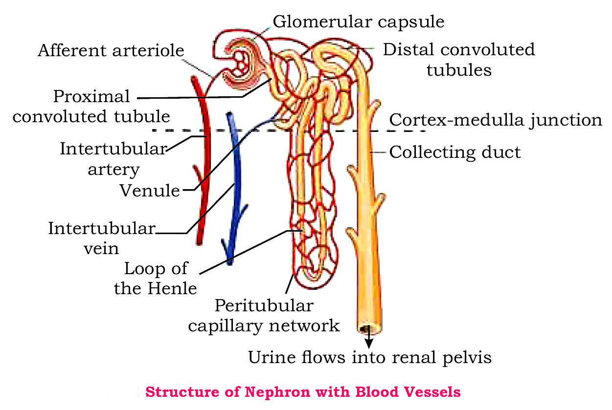
Parts of Nephron:
Each nephron is sub-divided into four main parts:
- Bowman's Capsule
- Proximal convoluted tubule
- Loop of Henle's
- Distal convoluted tubule
1. Bowman's Capsule:
- In each nephron inner end forms a cup-shaped swelling called Bowman's capsule.
- Each bowman's capsule have a ball of capillaries called glomerulus.
- Bowman's capsule with glomerulus are collectively called Malpighian body or Renal corpuscle.
- Bowman capsule leads into a short convoluted (coiled) tubule called Proximal Convoluted Tubule which passes into the medulla.
- The tubule enters into medulla, extends into renal pyramid and makes a U shaped structure called loop of Henle and it goes back into cortex.
- When tubule enters the cortex again, it becomes convoluted again.
- Function:
At the distal convoluted tubule, some water and minerals salts are reabsorbed.
-
Number of nephrons opens into a tube called collecting duct.
- Nephrons are surrounded by different blood vessels that are connected to the renal artery and renal vein.
STRUCTURE OF NEPHRON
OR

Q.20: Describe the function of nephrons? OR Describe the network of blood vessels found in nephron and their functions?
Ans: Function Of Nephrons OR Network And Functions of Blood Vessels In Nephron:
Nephrons remove waste materials and excess water from blood and convert them into urine by the process of filtration, reabsorption, secretion and excretion. Nephrons are surrounded by different blood vessels that are connected to the renal artery and renal vein. The blood filter to produce urine in blood capillaries of nephrons as:
- Afferent Arteriole:
The renal artery when enters into kidney, it divides into millions of branches called afferent arteriole.
Function: The blood enters the kidney through renal artery and goes into afferent arteriole. - Malpighian Body:
Each afferent arteriole further divides into numerous blood capillaries in Bowman's capsule are collectively called glomerulus. The Bowman's capsule with glomerulus is called Malpighian body or renal corpuscles.
Function: Ultrafiltration occurs at Malpighian body. - Efferent Arteriole:
Blood leaving the glomerulus through efferent arteriole, enter in blood capillaries surrounding the nephrons.
Function: these blood vessels carry away filtered blood from glomerulus. - Venule:
Blood capillaries surround a loop of Henle's unite to form venule, which ultimately joins to form a branch of renal vein.
Function: It carries filtered blood with less waste materials to the venous system.
Q.21: Draw labelled diagram of:
- Bowman's capsule and
- Section of kidney showing structure of two nephrons with blood supply.
BOWMAN'S CAPSULE

SECTION OF KIDNEY SHOWING STRUCTURE OF TWO NEPHRONS WITH BLOOD SUPPLY

OR

Q.22: Describe the role of Kidney in urine formation?
Ans: Role Of Kidney In Urine Formation:
Urine formation takes place in the following steps:
Urea formation:
The urea is formed with in the liver cells when the liver breaks down protein or amino acids, and ammonia.
The liver stores surplus glucose of food by converting it into glycogen and other food substances but it cannot store protein. The excess amino acids break and give some energy from it. The amino group (-NH2) is removed from amino acid called deamination and is converted into ammonia (NH3) which is very poisonous, it may kill the cell when stored in high concentration. So the liver cells quickly convert NH3 into less toxic substance urea. This urea is carried by blood to kidneys and excreted out in the form of urine. A small amount of urea is also excreted in sweat as well.
Urine formation:
URINE:
The mixture of excessive mineral salts and nitrogenous waste products i.e. urea, creatinine and uric acid (which are poisonous if accumulated), These are removed from body with water. This mixture is called urine.
Urine formation takes place in the kidneys. The two main processes are involved in the formation of urine within nephrons, which are:
- Filtration
- Reabsorption
(i) Filtration:
The process of taking out materials from blood is called filtration . It is of two types:
- (a) Ultrafiltration
- (b) Selective filtration
(a) Ultrafiltration:
Ultrafiltration occurs at Malpighian body when the blood from afferent arteriole enter into glomerulus located in Bowman's capsule. Most of the blood plasma is forced out of the glomerulus blood capillaries onto Bowman's capsule without any selection. This Process of non-selective filtration is called ultrafiltration.

(b) Selective filtration:
It occurs at proximal and distal convoluted tubules when blood flows into peritubular capillaries, the remaining amount of urea filters out from blood by active transport. It requires some energy.
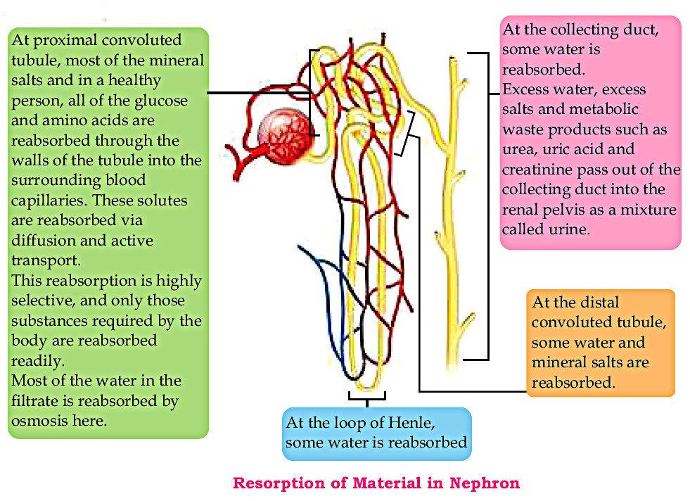
(ii) Reabsorption:
In a normal adult about 120 cm3 of filtrate is formed in the kidney every minute. If this large amount of filtrate allowed to passing out from the body as urine, the body will dehydrate and death may occur. To prevent this huge loss of water and useful salts, when the filtrate passes through the nephron useful substances and excessive water are reabsorbed in blood stream by:
- (a) Non-selective
- (b) Selective reabsorption
(a) Non-selective reabsorption:
It occurs at distal and proximal convoluted tubules without any selection.
- At the distal convoluted tubule, some water and minerals salts are reabsorbed.
- At proximal convoluted tubule, most the mineral salts and in a healthy person, all of the glucose and amino acids are reabsorbed through the walls of the tubules into the surrounding blood capillaries. These solutes are absorbed via diffusion and active transport.
This absorption is highly selective in quantity, and only those substances required by the body are reabsorbed readily.
Most of the eater in the filtrate is reabsorbed by osmosis here.
(b) Selective reabsorption:
It occurs at Loop of Henle's and collecting duct with the help of hormones. i.e. Antidiuretic hormone (ADH), Parathyroid hormones (PTH) and calcitonin.
- At the loop of Henle, some water is reabsorbed.
- At the collecting duct, some water is reabsorbed. Excess water, excess salts and metabolic waste products such as urea, uric acid and creatinine pass out of the collecting duct into renal pelvis as a mixture called urine.
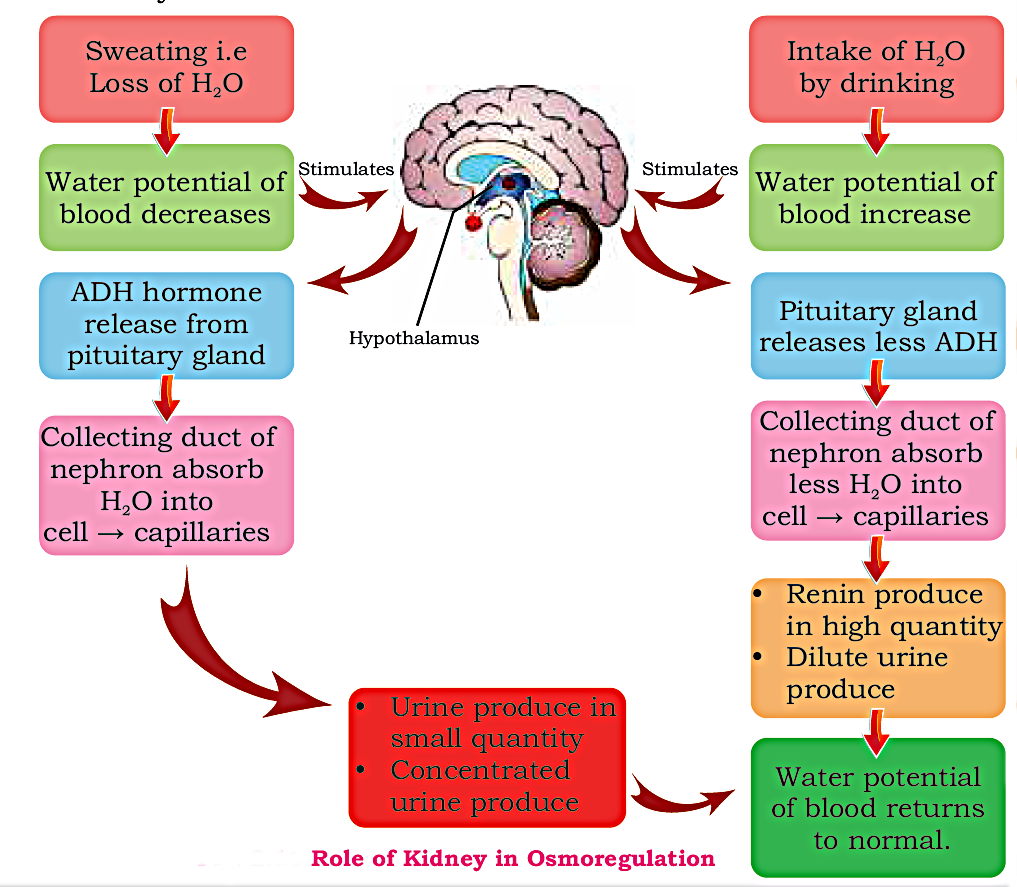
Q.23: Describe the role of kidneys in osmoregulation?
Ans: Role of kidneys in osmoregulation:
- The maintenance of internal water and salt conditions of the body by osmosis is known as osmoregulation.
- The water potential (capacity to lose water) of blood in the body has to be kept constant because big and sudden change in the water potential of blood can lead to serious problems.
e.g. if plasma becomes very much dilute water will enter the blood cells will swell and possibly burst. - On the other hand if the blood plasma becomes too concentrated, water will move out of the cell by osmosis, as a result of it the blood cells and tissues will become dehydrated and shrink. This control of water and salt content of the body is known as osmoregulation.
- Kidney is not only an excretory organ; it also regulates water and salt balance in the blood. Kidney ensures that concentration of blood stays more or less constant.
Q.24: Describe different disorders of kidney and their treatment?
Ans: Kidney Problems Or Disease:
When the kidneys do not function properly due to different reasons this is referred as Kidney problem or Kidney disease.
There are many problems of kidney:
- Kidney Stone
- Kidney Failure
Kidney Stone
A kidney stone is a solid mass that forms from the crystals of calcium oxalate or Calcium Carbonate. Sometimes uric acid and cysteine are also present in it. These molecules separate from urine, precipitate in kidney and deposit in the form of stone. Sometimes these stones are not hard therefore they break into sand like crystals which can pass out of the body with urine without pain. The little large size stone however damages the kidney tissues; it may stuck anywhere in urinary tract and cause renal failure with pain.
Treatment:
- Lithotripsy:
If the size of stone is comparatively small we can use the technique of lithotripsy to break stone by ultrasonic waves (sound waves). The broken rudiments drain out from kidney with urine. - Renal surgery:
The large size stone cannot be broken by lithotripsy, so it is removed only by the process of renal surgery.
Prevention:
The large intake of water is the only measure to minimize the chances of formation of stone in kidney.

Kidney failure:
Sometimes the nephrons of kidney are badly damaged and stop working due to certain reason or infection and the kidneys are not able to filter the harmful nitrogenous substance it is called Renal failure Or Kidney failure.
It is mainly due to solute dis-balance in blood and kidneys. The failure of kidneys allow urea and other waste materials to accumulate in blood. The amount of H2O is not regulated also. This dis-balance of solutes causes death unless the patient is given treatment to filter out wastes by machines.
Treatment of Kidney Failure:
- Kidney Transplant:
Patient of kidney failure may get a kidney transplant. In high degree renal failure the surgical transplantation of matching donor kidney is only the option left as the permanent treatment. A person with two healthy kidneys may donate one kidney and survive with one kidney. -
Dialysis:
Dialysis machine: A dialysis machine performs the function of a kidney. It helps to clean the patient's blood from metabolic waste products and toxic.
If a donor is not available, the patient can be treated with dialysis using a dialysis machine. For effective treatment the patient needs to undergo dialysis 2-3 times a week. Each session lasts about 3-5 hours depending on the patient's body size and medical condition.

Q.25: Differentiate between:
(i) Thermoregulation in hot condition and thermoregulation in cold condition.
(ii) Vasodialtion and vasocontraction.
Ans: (i) Difference Between Thermoregulation In Hot Condition And Thermoregulation In Cold Condition
| S.NO. | Thermoregulation In Hot Condition |
Thermoregulation In Cold Condition |
|---|---|---|
| 1. | In it body temperature rises. | In it body temperature falls. |
| 2. | Sweat glands start producing and secreting sweats. | Sweat glands stop to produce and secrete sweats. |
| 3. | Heat is lost to the environment. | Heat is retain. |
| 4. | Vasodilation occurs i.e. blood vessels dialte resulting in the heat lost to the environment. | Vasocontraction occurs i.e. blood vessels contract resulting in the heat is conserved. |
| 5. | Muscles attach with hairs are relaxed, causing laying down of hairs. | Muscles attach with hairs are contracted, pulling the hairs upright (erect). |
| 6. | Metabolic rate is decreases. | Metabolic rate is increases. |
(ii) Difference Between Vasodilation And Vasocontraction
| S.NO. | Vasodilation | Vasocontraction |
|---|---|---|
| 1. | When blood vessels (arterioles) in the dermis of skin dilates (become widen), it is called vasodilation. | When blood vessels (arterioles) in the dermis of skin contracts (become narrowed), it is called vasocontraction. |
| 2. | This process occurs in hot condition (Summer). | This process occurs in cold condition (winter). |
| 3. | In hot condition, body temperature start rises, which increases the flow of blood in capillaries near the skin. | In cold condition, body temperature start decreasing, which reduces the flow of blood in capillaries near the skin. |
| 4. | Blood pressure inside the blood vessels decreases. | Blood pressure inside the blood vessels increases. |
| 5. | It allows more heat loss to maintain thermoregualtion. | ;It retain heat to maintain thermoregualtion. |
Source: Special Thanks To Sir Syed Arif Ali
Thanks you so much
ReplyDeleteIt help me so much